 positive
 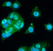 request CD with images
Cytoplasm to Nucleus Tanslocation Image Sets
1. Vitra plate
This 96-well plate has images of cytoplasm to nucleus translocation of the transcription factor NFκB in MCF7 and A549 cells in response to TNFα concentration. It has 12 concentration points in columns and 4 replica rows for each cell type. The plate was acquired at Vitra Bioscience on the CellCard reader - a microscope-based system at 10X, 4X, 2X objective magnification. For each well there is one field with two images - a nuclear counterstain (DAPI) image and a signal stain (FITC) image. Image size is 1360*1024 pixels. Images are in 8-bit BMP format. File name structure is <channel>-<well-number>-<row>-<column>-<field>.BMP
negative
positive
request CD with images
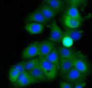
2. BioImage plate
This 96-well plate has images of cytoplasm to nucleus translocation of the Forkhead (FKHR-EGFP) fusion protein in stably transfected human osteosarcoma cells, U2OS. In proliferating cells FKHR is localized in the cytoplasm. Even without stimulation, Forkhead is constantly moving into the nucleus, but is transported out again by export proteins. Upon inhibition of nuclear export, FKHR accumulates in the nucleus. In this assay, export is inhibited by blocking PI3 kinase / PKB signaling by incubating cells for 1 hr with Wortmannin and with compound LY294002. There are 4 replicas of the 9-point dose curve for each drug. Nuclear counterstain is DRAQ. The images were acquired at BioImage on the IN Cell Analyzer 3000 and are available in native FRM format and converted to 8-bit BMP format with one image per channel. Image size is 640*640 pixels. File name structure in FRM format is <prefix>_<row><column>_<suffix>.frm; file name structure in BMP format is <channel>-<well-number>-<row>-<column>.BMP
negative 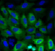 positive
 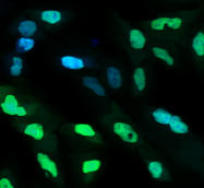 request CD with images